- +91-8052038976
- drrishishukla@gmail.com
- SWAROOP NAGAR, KANPUR 208002
Ophthalmic Services
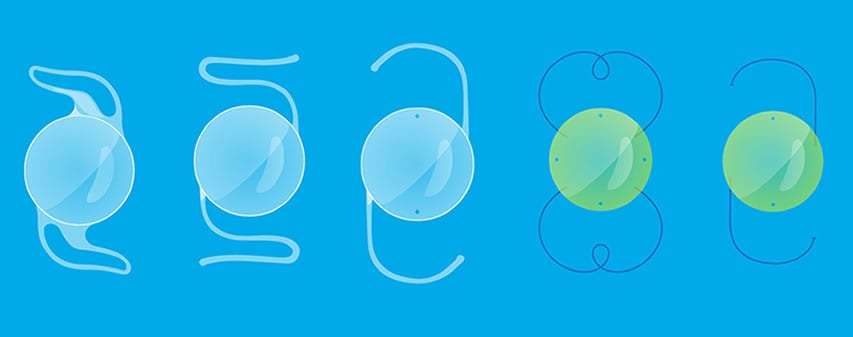
What are IOLs?
Intraocular lenses (IOLs) are medical devices that are implanted inside the eye to replace the eye’s natural lens when it is removed during cataract surgery. IOLs also are used for a type of vision correction surgery called refractive lens exchange.


Cataract Services Phacoemulsification with Premium IOLs
We specialize in treating the cataract through Phacoemulsification. It is a modern cataract surgery in which the eye’s internal lens is emulsified with an ultrasonic handpiece and aspirated from the eye. Aspirated fluids are replaced with Irrigation of balanced salt solution to maintain the anterior chamber.
Premium IOLs used in our Centre includes
Aspheric Monofocal IOLs, Toric IOLS, Multifocal IOLs, Trifocal IOLs and Extended Range of Vision IOLs (US FDA Approved).
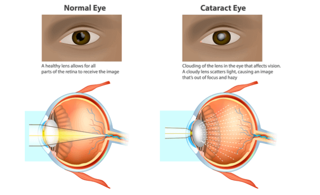
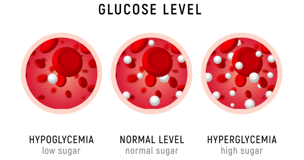
Specialized Type 2 Diabetes in Cataract
Cataract in diabetic patients is a major cause of blindness. The pathogenesis of diabetic cataract development is still not fülly understood. However,Phacoemulsification is nowadays the preferred technique in most types of cataract whether it has developed because of diabetes or just with old-ageOverall outcomes of cataract surgery are excellent.
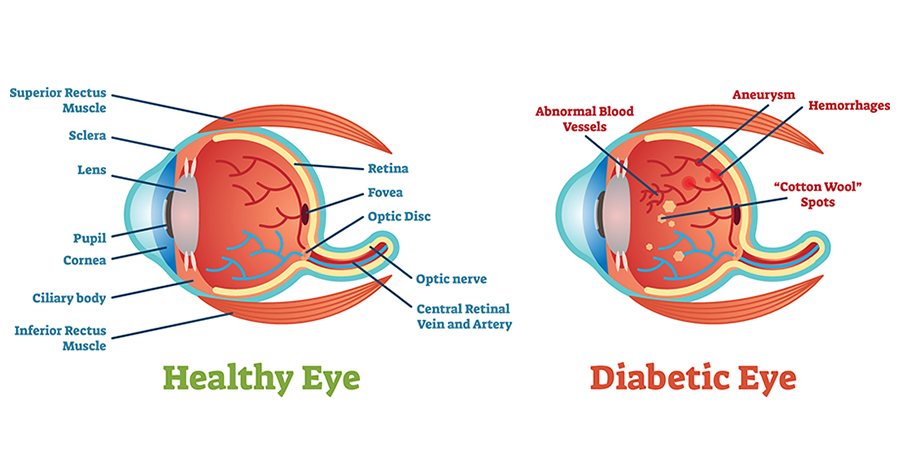
Most Experienced in Adolescent with Type 1 Diabetes
The prevalence of early diabetic cataract in children and adolescents with T1DM ranges between 0.7 % to 3.4 %. The occurrence of diabetic cataract in most pediatric patients is the first sign of T1DM or occurs within 6 months of diagnosis of T1DM. Today, there are many experimental therapies for the treatment of diabetic cataract, but cataract surgery continues to be a gold standard In the treatment of diabetic cataract. Since the cataract is the leading cause of visual Impairment in patients with T1DM, diabetic cataract requires an Initial screening as well as continuous surveillance as a measure of prevention. Our center specializes in dealing with vision problems occurring in adolescents with type 1 diabetes. A good number of cataract surgery with IOLimplantation in Type 1 diabetics have been performed successfully in the center.
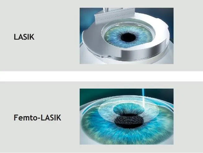
LASIK LASER (FEMTO & C-LASIK)
FEMTO – Lasik: Only Femto-Lasik Centre in Kanpur! Bladeless Lasik Surgery is the most advanced procedure being used by ophthalmologists today. Itis also known as Femto Lasik, blade-free Lasik or all-laser lasik. In this procedure Lasik surgeon uses two types of lasers for the Vision correction procedure. First, an ultra-fast Femto. Second laser is used to create a thin flap in the cornea. C-LASIK: The very latest development in laser eye treatment is called C Lasik, a system that takes LASIK to a new frontier. It is the fully integrated personalized correction system that helps patients achieves better quality of vision.

Yag Laser
A Yag capsulotomy is a special laser treatment used to improve your vision after cataract surgery. It is a simple, commonly performed procedure which is very safe. During your cataract operation, the natural lens inside your eye that had become cloudy was removed.
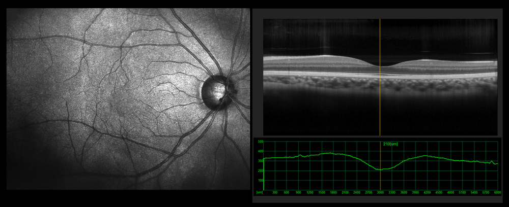
Retina Examination - Fundus Imaging. OCT
OCT: Optical Coherence Tomography (OCT) is a non-invasive imaging test. OCT uses light waves to take cross-section pictures of your retina. With OCT,your ophthalmologist can see each of the retina’s distinctive layers. It helps in detection of retinopathy and glaucoma.
Fundus Imaging: Fundus Imaging is used to inspect anomalies associated with diseases that affect the eye, and to monitor their progression. It is able to identify glaucoma and multiple sclerosis, as well as monitor disease processes such as macular degeneration, retinal neoplasms, choroid disturbances and diabetic retinopathy. Fundus Imaging assist in the planning of additional management options for these irregularities. The medical necessity of fundus must be recorded comprehensively so that the clinician is able to compare photographs of a patient from different timelines.

Refraction - ARK
Autorefractor Keretometer (ARK) is a computer-controlled machine used during an eye examination to provide an objective measurement of a person’s refractive error and prescription for glasses or contact lenses. This is achieved by measuring how light is changed as it enters a person’s eye. Like many ophthalmology instruments, they have spherical and cylindrical measurement ranges of-30 to +25D/±100 respectively.
Contact Lenses
A contact lens is a thin, curved lens placed on the film of tears that covers the surface of the eye. The lens itself is naturally clear but is often given the slightest tinge of color to make them easier for wearers to handle. Today’s contact lenses are either hard or soft.

Glaucoma Diagnosis and Management
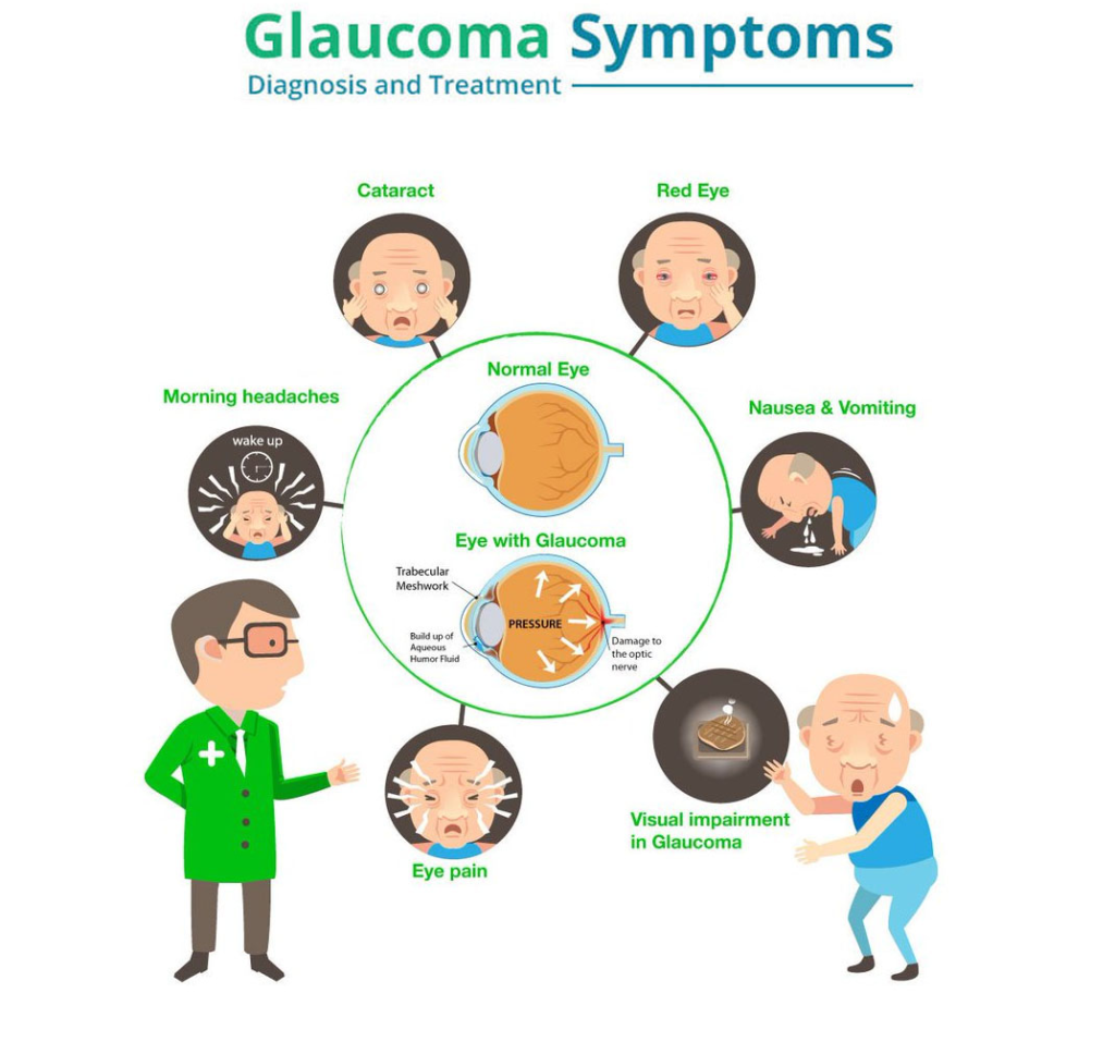
Glaucoma Diagnosis
- Measure Eye Pressure
- Inspect Eye’s Drainage Angle
- Examine Optic Nerve for Damage
- Test Peripheral (side) Vision
- Take a picture or Computer Measurement of Optic Nerve
- Measure the thickness of Cornea
Glaucoma Management: Glaucoma is treated by lowering your eye pressure (intraocular pressure). Depending on your situation, your options may include prescription eye drops, oral medications, laser treatment, surgery or a combination of any of these. Micro pulse laser is the latest treatment technique in Glaucoma.
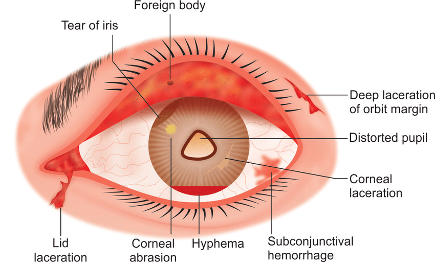
Ocular Trauma in Diabetics
Our center is fully equipped to treat any emergency situation in-case of eye injury in a diabetic patient.

NCT & Applanation Tonometry
NCT: It is a test used during an eye exam to measure the pressure Inside your eye. The air puff test gives your eye doctor an eye pressure reading, known as intraocular pressure (1OP), which helps detect glaucoma.
Applanation Tonometry: It is a test measures of pressure it takes to flatten a portion of your cornea, pressure readings help your doctor diagnose and keep track of glaucoma.
Orthoptics
Orthoptics is an ophthalmic field pertaining to the evaluation and treatment of patients with disorders of the visual system with an emphasis on binocular vision and eye movements. Orthoptists are uniquely skilled in diagnostic techniques.
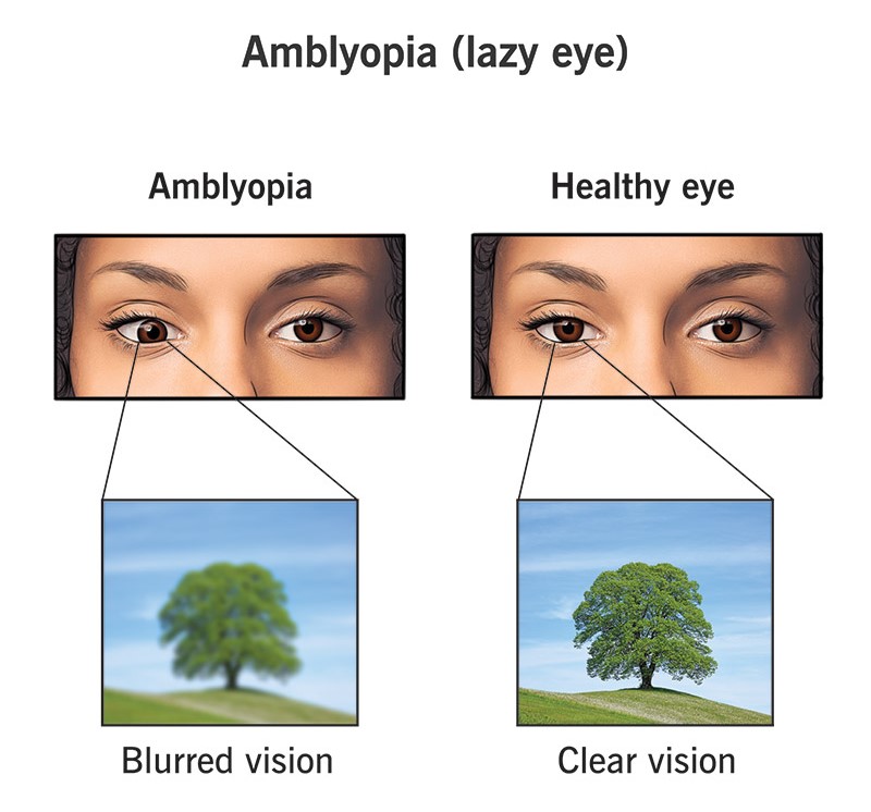
Amblyopic Management
Amblyopia treatment typically consists of refractive correction followed by part-time patching or atropine penalization of the fellow eye to promote use of the amblyopic eye. Six months to two years of patching results in improved function in a majority of cases.
Squint Management
- Eyeglasses or Contact Lenses: Used in patients with uncorrected refractive errors. With corrective lenses, the eyes will need less focusing effort and may remain straight.
- Prism Lenses: Special lenses that can bend light entering the eye and help reduce the amount of turning the eye must do to look at objects.
- Orthoptics (Eye Exercises): May work on some types of strabismus, especially convergence insufficiency(a form of exotropia).
- Medications: Eye drops or ointments. Also, Injections of botulinum toxin type A (such as Botox) can weaken an overactive eye muscle. These treatments may be used with, or in place of, surgery, depending on the patient’s situation.
- Patching: To treat amblyopia (lazy eye), if the patient has it at the same time as strabismus. The improvement of vision may also improve control of eye misalignment.
- Eye Muscle Surgery: Surgery changes the length or position of eye muscles so that the eyes are aligned correctly. This is performed under general anesthesia with dissolvable stitches. Sometimes adults are offered adjustable strabismus surgery, where the eye muscle positions are adjusted after surgery.

If you need urgent care, simply call our 24 hour emergency hotline.
Your personal case manager will ensure that you receive the best possible care.
Call Now